Class III Malocclusion:
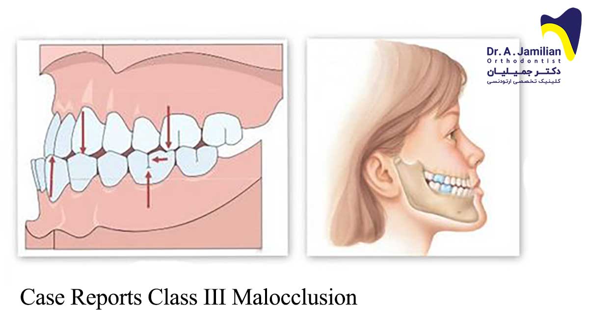
This case report demonstrates a 17.9-year-old boy with marked high angle skeletal class III malocclusion with severe maxillary retrognathia and mandibular prognathism.
Summary:
The patient had crowding in the upper jaw and the lower incisors were tipped lingually due to Class III malocclusion compensation. Class III molar and canine relationship with posterior cross bite. High maxillary-mandibular plane angle. Incompetent lips with strain.
Examination of head and face:
In the frontal plane the face of the patient has an elongated shape. Skeletal Class III pattern with severe maxillary retrusion and mandibular prognathism can be seen. Concave profile can also be seen. Incompetent lips with strain.
Functional examination:
The patient’s path of closure showed no deviation. Maximum jaw opening was normal at 49 mm. The patient did not complain from any TMJ signs or symptoms, and there were no clicks or pain present on opening and closing. No pain on palpation could be detected.
Intra-oral examination:
Severe Class III molar and canine relationship. Anterior and posterior cross bite could be detected. He had a reverse overjet of 9 mm. Crowding could also be seen in the upper jaw. Retroclined lower incisors. The patient had fair oral hygiene with some restorations.
Treatment plan:
The patient’s chief complaints were as following:
Severe maxillary deficiency
Severe mandibular prognathism
Crowding of upper arch
Considering the severity of the malocclusion, the underlying skeletal discrepancy, the age of the patient and his expectations a surgical-orthodontic approach was deemed.
The treatment plan was as following:
Levelling and aligning
Decrowding
Incisor decompensation by means of fixed appliance
Bimaxillary surgery involving a maxillary advancement and impaction, and a mandibular setback (bilateral sagittal split ramus osteotomy). Chin Augmentation.
Post-surgical orthodontics: Arch coordination, detailing, suitable interdigitation
Debonding
Retention
Treatment results:
After orthodontic and surgical procedures a bilateral Class 1 molar and canine relationship has been achieved. Ideal overjet and overbite was also achieved. The patient has a more balanced Class 1 profile with good facial harmony and the face is totally symmetrical. The upper and lower dental midlines are coincident and match with the skeletal base. No signs or symptoms of temporomandibular joint dysfunction can be detected. Upper and lower Hawley appliances were placed in the jaws in order to ensure adequate retention.
The retainers were worn on a full time basis for the first 6 months then on a part time basis after which their use was discontinued. The patient returned to the office twice a year for orthodontic check. The patient was referred to an oral surgeon for extraction of his third molars.
Retention:
13 months after retention no change had occurred in the profile and occlusion of the patient. And no maxillary deficiency and jaw discrepancy can be seen. Intra oral examination showed that the patient is in molar and canine Class I relationship and there is no discrepancy between the jaws. The intercuspation was still satisfactory and no signs of bruxism or other dysfunction could be detected. The lips are competent and the patient is very satisfied with his appearance. No clicking or no signs and symptoms of temporomandibular dysfunction were noted.
Cephalometric data:
| Pretreatment | Posttreatment | Mean SD | ||
| Sagittal Skeletal Relations | ||||
| Maxillary Position S-N-A | 76° | 82° | 82º ± 3.5º | |
| Mandibular Position S-N-Pg | 82° | 80° | 80º ± 3.5º | |
| Sagittal Jaw Relation A-N-Pg | -6° | 2° | 2º ± 2.5º | |
| Vertical Skeletal Relations | ||||
| Maxillary Inclination S-N / ANS-PNS | 10° | 10° | 8º ± 3.0º | |
| Mandibular Inclination S-N / Go-Gn | 43° | 40° | 33º ± 2.5º | |
| Vertical Jaw Relation ANS-PNS / Go-Gn | 33° | 30° | 25º ± 6.0º | |
| Dento-Basal Relations | ||||
| Maxillary Incisor Inclination 1 – ANS-PNS | 107° | 113° | 110º ± 6.0º | |
| Mandibular Incisor Inclination 1 – Go-Gn | 71° | 84° | 94º ± 7.0º | |
| Mandibular Incisor Compensation 1 – A-Pg (mm) | 8 mm | 2 mm | 2 ± 2.0 | |
| Dental Relations | ||||
| Overjet (mm) | – 9 mm | 2 mm | 3.5 ± 2.5 | |
| Overbite (mm) | -2 mm | 2 mm | 2 ± 2.5 | |
| Interincisal Angle | 148 ° | 135° | 132º ± 6.0º |
The following images show the pre and post treatment lateral cephalogram and OPG of the patient:


