Case Reports-Arch Length Deficiency
Arch Length Deficiency
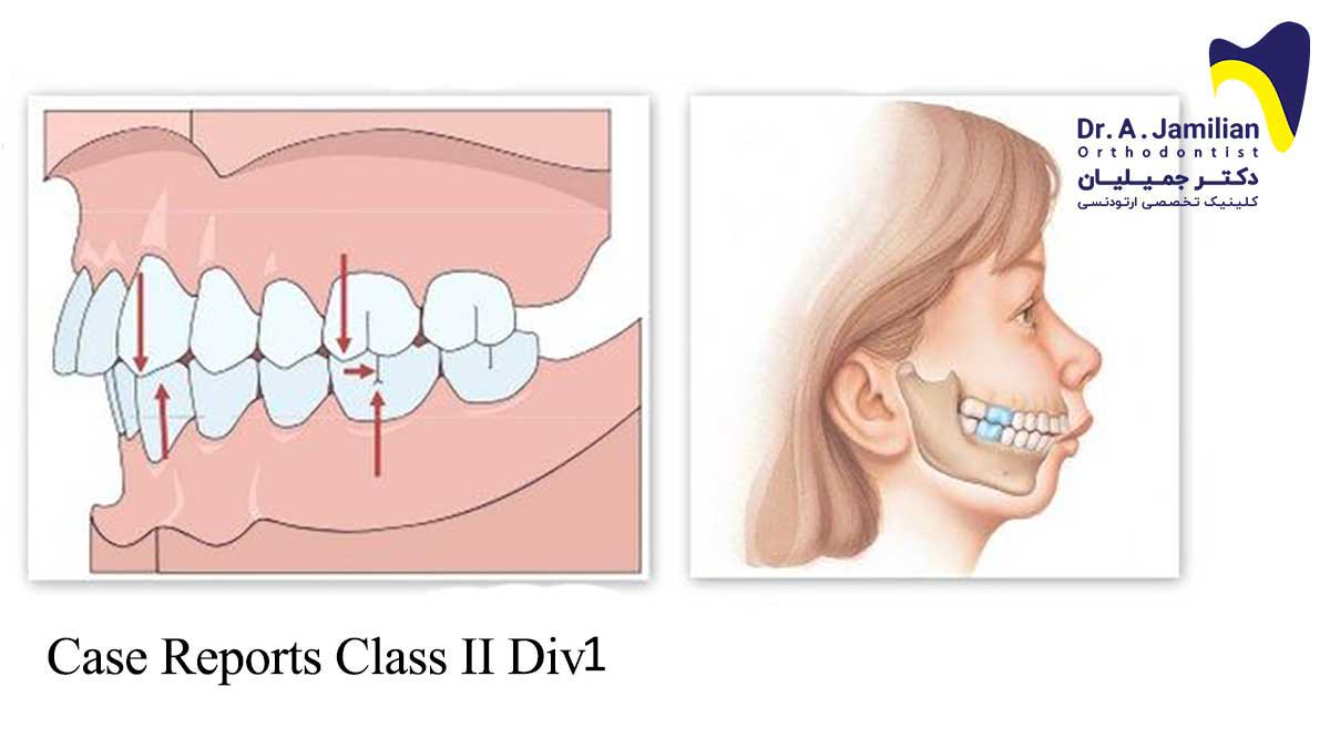
This case report demonstrates a 9.4-year-old boy in the mixed dentition presenting with arch length deficiency along with skeletal Class II Division 1 high angle malocclusion (Sn-GoGn=45°).
Summary:
The patient had severe mandibular deficiency with increased anterior facial height. Severly flared lower incisors. No facial assymetry. Incompetent lips.
Examination of head and face:
In the frontal plane the face of the patient had an elongated shape with vertical growth pattern. In the sagittal plane the profile shows convexity with severe mandibular deficiency. Lips were incompetent. No facial asymmetry could be seen.
Functional examination:
The patient did not complain from any TMJ signs or symptoms, and there were no clicks or pain present on opening and closing. No lateral mandibular shift could be assessed during examination. Centric occlusion and centric relation were coincident.
Intra-oral examination:
The patient is in mixed dentition. The molars and canines were in Class 1 relationship. Increased overjet was masked by severely flared lower incisors. He had generalized gingival enlargement which could also be seen in his father. The patient had well aligned arches in both jaws. 6 of his teeth had restorations which was indicative of poor oral hygiene.
Treatment plan:
The chief concerns were as following:
Class II Div 1 malocclusion with severe maxillo mandibular discrepancy
Mandibular deficiency
Chin retrognathia
Lip incompetency
Flared lower incisors
Avoiding invasive surgical procedures
Severe maxillo mandibular discrepancy and high mandibular plane angle made this patient a candidate for surgical correction. However, growth modification was thought to be more appropriate to treat Class II relationship and achieve skeletal and dental changes. Mandibular deficiency and lack of overjet made the treatment of this patient quite challenging.
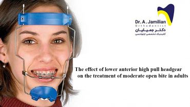
Use of functional appliance requires enough overjet. An Upper removable appliance would flare the upper incisors and create the required overjet. Afterwards, for growth modification, it was decided to use a Twin block functional appliance in conjunction with high-pull headgear. High-pull headgear would impact posterior section of the maxilla which would allow autorotation of the mandible and Twin block would move the mandible forward. In the meantime, lower deciduous canines and first & second molars would be extracted. These extractions would allow earlier eruption of permanent first premolars. Afterward, first premolars would also be extracted in order to reduce the flaring of lower incisors. Extraction of upper and lower first premolars would also help to reduce vertical dimension of the patient. The treatment could be continued by fixed orthodontics.
Treatment results:
The patient’s Class II profile has improved due to skeletal and dentoalveolar changes. There has been a reduction in the sagittal discrepancy and mandibular retrognathia due to the enhancement of mandibular forward growth and development.
The patient’s vertical pattern improved significantly and it changed to normal growth pattern. Thus, his facial height reduced favourably. Anterior upper and lower teeth inclination changed from flared to upright position. The final interdigitation is satisfactory. The lips became competent and the patient’s aesthetics became satisfactory. Normal overjet and overbite has also been established. Total treatment time was 32 months during which the Class II division 1 profile has been improved. The incompetent lips were also corrected and there was no more sign of incompetent lips. Since the patient had tendency towards vertical growth pattern, posterior bit plane was chosen to prevent any relapse and lower Hawley retainer was used to ensure adequate retention. The retainers were worn on a full time basis for the first 6 months and then on a part time basis for a further 6 months after which their use was discontinued.
Retention:
13 months out of retention the patient’s profile was satisfactory and there was no sign of mandibular retrognathia, flaring of lower incisors, and vertical growth pattern. His occlusion was satisfactory. No relapse could be seen in the post retention records. The temporomandibular joint showed no clicking on opening or closing. No deviation could be detected upon opening of the jaws.
Cephalometric Data:
| Pretreatment | Posttreatment | Mean SD | ||
| Sagittal Skeletal Relations | ||||
| Maxillary Position S-N-A | 85° | 80° | 82º ± 3.5º | |
| Mandibular Position S-N-Pg | 73° | 76° | 80º ± 3.5º | |
| Sagittal Jaw Relation A-N-Pg | 12° | 4° | 2º ± 2.5º | |
| Vertical Skeletal Relations | ||||
| Maxillary Inclination S-N / ANS-PNS | 6° | 9° | 8º ± 3.0º | |
| Mandibular Inclination S-N / Go-Gn | 45° | 40° | 33º ± 2.5º | |
| Vertical Jaw Relation ANS-PNS / Go-Gn | 41° | 32° | 25º ± 6.0º | |
| Dento-Basal Relations | ||||
| Maxillary Incisor Inclination 1 – ANS-PNS | 108° | 98° | 110º ± 6.0º | |
| Mandibular Incisor Inclination 1 – Go-Gn | 102° | 94° | 94º ± 7.0º | |
| Mandibular Incisor Compensation 1 – A-Pg (mm) | 10 mm | 3 mm | 2 ± 2.0 | |
| Dental Relations | ||||
| Overjet (mm) | 5 mm | 2 mm | 3.5 ± 2.5 | |
| Overbite (mm) | 1 mm | 2 mm | 2 ± 2.5 | |
| Interincisal Angle | 110° | 138° | 132º ± 6.0º |
The following images show the pre and post treatment lateral cephalogram and OPG of the patient:


