Dental radiography is a diagnostic imaging technique that uses X-rays to generate detailed images of the teeth and mouth. These images enable dentists to detect dental issues, e.g., cavities, gum disease, root canal problems, and infections that might be invisible during a routine examination. Dental X-rays facilitate the quick and accurate diagnosis of dental problems and thus prevent more serious issues from developing. Therefore, dental radiography is crucial for making treatment plans, monitoring treatment progress, and assessing the overall condition of a patient’s jaw and teeth. Dental radiography can be categorized into two main types: intraoral and extraoral imaging. Below, we describe each of these methods in full detail.
1. Intraoral Dental Radiology
Intraoral dental radiography provides detailed images of your teeth and surrounding structures by placing a small film or digital sensor inside your mouth. By capturing high-quality images of small dental areas, intraoral radiography enables dentists to detect a wide range of dental issues, e.g., cavities, changes in tooth structure, gum disease, and impacted or misaligned teeth. These conditions are typically invisible during a standard dental examination and can only be identified through dental X-rays. The most common intraoral radiography techniques include:
1.1. Bitewing X-Rays
A bitewing X-ray is a common type of dental X-ray that provides a detailed view of a tooth from the crown to the level of the bone that supports it. Dentists use bitewing X-rays to detect cavities between teeth and changes in bone thickness caused by gum disease. During bitewing radiography, a small film is placed in your mouth, and you bite down on its paper tab to hold it in place. An X-ray machine is then positioned over your teeth to capture an image. Digital images are available instantly, and the radiographic film is developed within a few minutes. To protect your body from radiation, you will be given a lead apron to cover your chest and abdomen. While the radiographic radiation dose from a bitewing X-ray is minimal, limiting unnecessary exposure is essential. Bitewing X-rays are valuable for assessing gum health and the bone structure around your teeth, and they are often used during periodic dental checkups to identify cavities between teeth. Additionally, bitewing X-rays can be taken of baby teeth to monitor the development of permanent teeth in children.
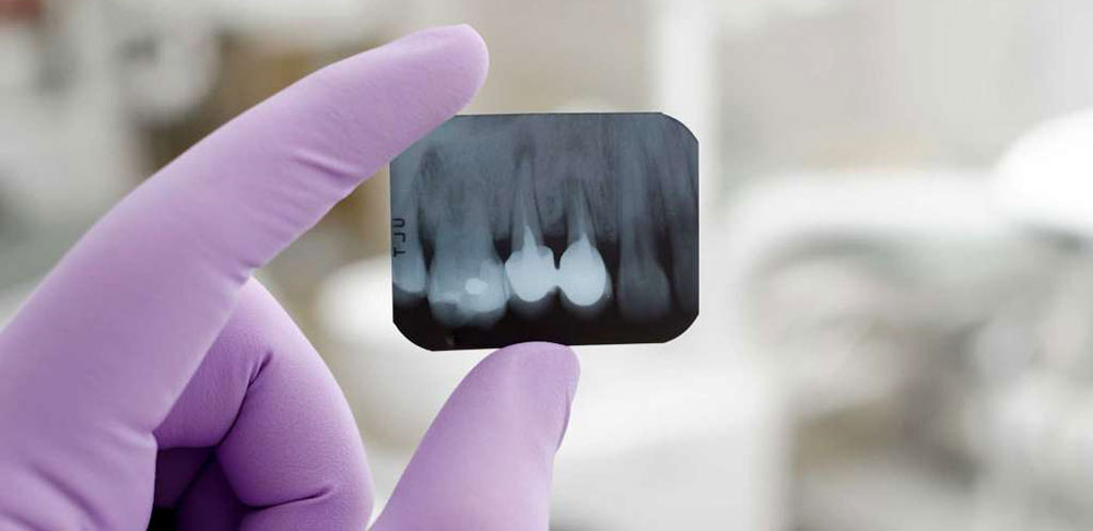
1.2. Periapical Radiography
Periapical radiography is a precise dental imaging technique that captures detailed images of a specific tooth and its surrounding areas. By directing X-rays onto the tooth and its root, dentists can effectively detect various dental issues, e.g., cavities, fractures, infections, and potential tumors. Commonly, periapical X-rays capture images of one to three teeth at a time. These images help diagnose decay, especially in the chewing surfaces of the teeth. Additionally, periapical radiography plays a crucial role in root canal treatments. Dentists often take an initial X-ray to assess the tooth root before beginning treatment. Subsequent X-rays are taken throughout the procedure to monitor the length and placement of dental files or needles within the root canal. The third and fourth periapical X-rays ensure that the root canal is filled and the treatment is successful.
1.3. Occlusal Radiography
Occlusal radiography is a dental imaging technique that obtains an occlusal view of the upper or lower jaw. During this procedure, the patient positions their teeth specifically to allow for a clear image of the mouth’s internal structures. This diagnostic tool is invaluable for detecting a wide range of oral conditions, e.g., bone injuries, tumors, cysts, and other abnormalities. Occlusal X-rays are frequently employed when a more comprehensive and accurate view of the jaw or teeth is required.
2. Extraoral Dental Radiology
Extraoral radiography involves positioning the film or sensor outside the mouth. This method provides more comprehensive views of teeth, jaws, sinuses, and facial bones. It is an advanced diagnostic tool in dentistry for detecting dental and oral structural issues. Common extra-oral radiography techniques include:
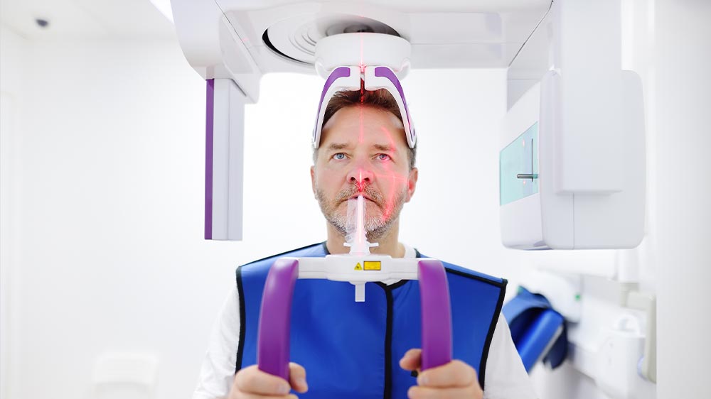
2.1. Panoramic Dental Radiology Orthopantomogram or OPG)
A panoramic X-ray, also known as an OPG (i.e. orthopantomography), provides a wide view of all the teeth, jaws, surrounding bone structures, and other oral structures. An OPG is commonly used to assess dental health before surgical procedures and to diagnose widespread infections or tumors. During an OPG, a machine rotates around your head to capture a detailed image of your entire mouth. OPGs are invaluable for diagnosing jaw fractures from accidents or falls, identifying jaw tumors and cysts, and evaluating dental and jaw conditions before orthodontic treatment. Dentists also rely on OPGs to assess the position and growth of wisdom teeth before extraction. While this imaging method offers numerous benefits, weighing the advantages against potential drawbacks is essential to understanding the true value. The merits and demerits of this radiological imaging technique are as follows:
- Merits: It provides a comprehensive image of all teeth and their surrounding structures, aids in diagnosing broad problems, and is faster to perform than other methods.
- Demerits: Finer details of dental problems, particularly in specific areas such as the roots, may not be visible in the captured image, and images may be taken at a lower resolution than other methods.
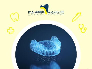
2.2. Cone-Beam Computed Tomography (CBCT)
CBCT is an advanced imaging technique that provides cross-sectional, 3D images of oral structures. CBCT scans allow dentists to identify problems that are not visible on panoramic or other radiological images. This advanced imaging is invaluable for examining a wide range of dental conditions, e.g., jaw bones and dental implants, diagnosing infections, or dental disorders. Dentists commonly use CBCT scans to examine the position of the jaw and teeth with high clarity and accuracy before they plan dental implant procedures:
- Merits: High diagnostic accuracy for complex bone and jaw problems, as well as cross-sectional images with precise and practical details in implants
- ·Demerits: More expensive than other imaging methods; specialized equipment needed
2.3. Cephalometric Radiography
Cephalometric radiography provides images of the face and oral structures, e.g., the jaws and teeth. This specialized imaging technique is extensively used in orthodontics to assess the growth of the jaws and teeth and to plan orthodontic treatment. Cephalometric images are particularly valuable in orthodontics as they enable the prediction of structural changes in the jaws and face during treatment.
- Merits: It is used for treatment planning at the beginning of orthodontic treatment or jaw surgery. It helps evaluate jaw changes during orthodontic treatments to ensure more accurate treatment planning.
- Demerits: This procedure requires special equipment and is only useful for diagnosing jaw and facial problems.
Ensure that your dental X-rays are taken at offices equipped with various types of radiology equipment. If an orthodontic treatment is to be underway, X-rays are typically essential. These specialized images are particularly useful for assessing jaw, face, and skull growth patterns. They also help determine jaw and face growth direction and facial pattern. An orthodontic treatment requires a certain cephalometric image known as lateral cephalometry.
2.4. Sialogram Radiography
Sialography is an imaging technique for evaluating the salivary glands, particularly when there is a suspicion of infection or blockage of the salivary ducts. This procedure involves injecting a contrast medium into the salivary ducts, followed by X-ray imaging.
- Merits: Highly effective in identifying issues related to the salivary glands, including blocked or inflamed salivary ducts·
- Demerits: It requires contrast medium injections. Some patients cannot tolerate these injections, which may cause minor side effects.
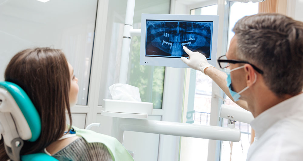
2.5. Digital Dental Radiography
Digital dental radiography is an advanced imaging technique that uses digital sensors instead of traditional X-ray films. It allows for immediate capture and electronic transmission of high-resolution dental images. Dentists can instantly and easily view these high-resolution images on a computer screen.
- Merits: Quality images and quick completion; easy image storage and sharing.
- Demerits: Requires advanced and expensive digital equipment, which may not be available in all offices
Comparing the Merits and Demerits of Dental Radiology Techniques
The following table compares the merits and demerits of common extraoral radiology techniques:
| Extraoral Technique | Merits | Demerits |
|---|---|---|
| Panoramic (OPG) | 1. A comprehensive view of the teeth and their surrounding structure 2. Less time needed for imaging | 1. Failure to record precise details of specific areas, such as dental roots 2. Images have relatively low resolution and quality. 3. Exposure to radiation |
| Computed Tomography (CBCT) | 1. High diagnostic accuracy for complex bone and jaw problems 2. Offering cross-sectional images with precise and practical details in implants | 1. More expensive than other imaging methods. 2. Special equipment needed 3. Exposure to radiation |
| Cephalometric | 1. Suitable for orthodontic treatment 2. Appropriate for jaw surgery and assessment of jaw changes | 1. Special equipment needed 2. Limited to diagnosing jaw and facial complexities 3. Exposure to radiation |
| Sialogram | 1. Useful in identifying salivary gland issues, such as blocked or inflamed salivary ducts | 1. Contrast medium injected, which some patients cannot tolerate and may have minor side effects 2. Exposure to radiation |
| Digital | 1. High-quality images and less time to complete 2. Images that can be easily saved and shared | 1. Advanced and expensive digital equipment 2. Exposure to radiation |
The following table compares the merits and demerits of common intraoral radiology techniques:
| Intraoral Technique | Merits | Demerits |
|---|---|---|
| Bitewing | 1. Displays the tooth from crown to supporting bone surface 2. Examines the health of the gums and bone structure around the teeth | 1. Some diagnostic limitations 2. Patient cooperation needed 3. Exposure to radiation |
| Periapical | 1. Imaging a tooth and its surrounding areas | 1. Restrictions in specific areas of the teeth 2. Image quality reliant on the operator’s experience and skill 3. Exposure to radiation |
| Occlusal | 1. Ideal for diagnosing bone injuries, tumors, cysts, and other disorders | 1. Limitations in image detail 2. Limited diagnostic capacity for certain diseases 3. Patient positioning and angulation 4. Exposure to radiation |
How to Take a Dental X-Ray?
No special preparation is typically required if you want to take dental X-rays. Simply brush your teeth thoroughly before your appointment. You will sit in a dental X-ray chair and wear a lead apron to protect your body from radiation during the procedure. The lead apron is placed over sensitive areas such as your abdomen and chest to protect your internal organs.
If an intraoral X-ray is required, you will be given a plastic holder to bite down on. This holder keeps your teeth and jaw stable, resulting in clear images of your teeth, gums, and surrounding structures. The dental technician will position the X-ray machine at different angles to capture detailed images of all your teeth.
If you require an extraoral X-ray, the X-ray machine is set to take images of your teeth and jaw from outside your mouth. There is no need for you to bite into a bite block. The machine is positioned near your head to capture images of various areas of your mouth and teeth.
In this article at Dr. Jamilian’s website, we have looked at the different types of dental X-rays and how they are used to capture detailed images of your teeth. Your dentist will usually recommend a specific type of X-ray based on your individual dental needs and treatment plan. Each type of dental X-ray serves a specific diagnostic purpose, allowing dentists to make more informed decisions about your oral health.
FAQ about Dental Radiology
An OPG (orthopantomogram) scan is a 2D imaging technique that captures an image of the entire mouth, teeth, and jaws in a single scan. It is useful for diagnosing problems with tooth position, teeth growth and development, the jaw, and the temporomandibular joint (TMJ), as well as orthodontic treatment planning.
An OPG is a 2D imaging scan that gives a broad view of the mouth, whereas a CBCT is a 3D imaging scan that shows detailed, cross-sectional images of the teeth and jaws.
Due to the low radiation dose, the potential complications of dental radiology are usually minimal. It is best to provide the patient with adequate protection, e.g., a lead apron, during radiology.
Dentists analyze dental radiology images. Reading dental radiographs allows them to detect decay, root infections, impacted teeth, and fractures.
Pediatric dental radiology uses specialized equipment to minimize radiation exposure. Complications are usually minor; however, protective equipment, e.g., lead aprons, should always be used.
An X-ray of a decayed tooth reveals the extent of the decay and its precise location. The dentist uses the images to determine whether the tooth should be repaired, root canaled, or extracted.